
4D Ultrasound Is Changing How We Monitor Fetal Development
Three trimesters. 40 weeks. 280 days. No matter how you count it, childbirth doesn’t happen overnight. There’s an incredible amount of fetal development during gestation, and it’s crucial for moms and clinicians to follow fetal progress closely.
In 2022, typical pregnancy care includes a minimum of two ultrasound examinations: one to confirm the due date and the other to confirm normal fetal development. But potential complications can quickly make ultrasound imaging a more frequent occurrence. In these cases, 4D ultrasound technology is the standard of care for monitoring complications as they develop and informing treatment throughout pregnancy and postpartum care.
3D vs. 4D ultrasound
Ultrasound imaging has come a long way in the last 30 years. Black and white 2D ultrasounds, which are still common today, provide static images of a fetus in utero. Now, 3D and 4D ultrasound technology gives expectant parents and medical professionals a detailed look at fetal development. What’s the difference?
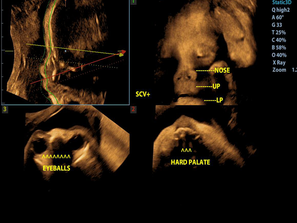
3D ultrasound allows doctors to examine internal and external fetal anatomy via static 3D images. 4D ultrasound takes the technology a step further. During a 4D ultrasound, the clinician can livestream video of anatomical images. This allows healthcare providers to observe the function of the fetal heart wall and valves, including blood flow through various vessels.
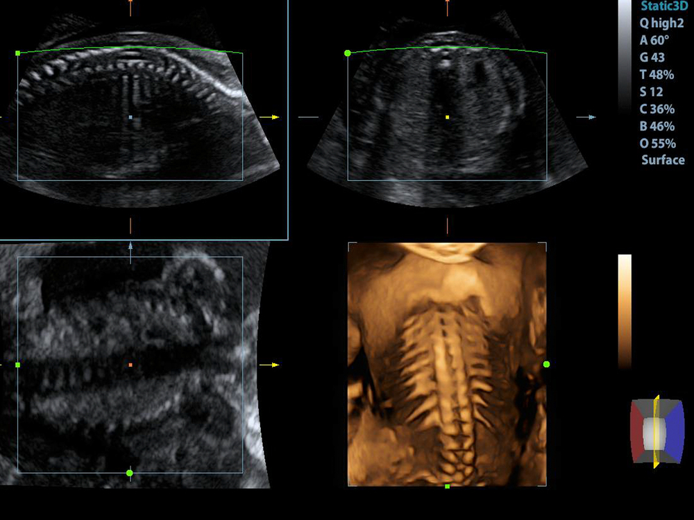
3D ultrasound uses several different imaging techniques to take 2D images of the fetus. Software merges the images to provide a 3D perspective of development in utero. Physicians can adjust reference points within the image to rotate the perspective.
4D ultrasound offers the same benefits in real time. Because 4D ultrasound produces live views of the fetus in motion, it is particularly useful for monitoring the development and function of internal anatomy.
 Advantages of 4D ultrasound technology
 Advantages of 4D ultrasound technology
The ability to stream real-time fetal development diagnostics provides significant advantages. 4D ultrasound is especially critical for high-risk pregnancies.
Trained healthcare providers use 4D ultrasound for:
- Enhanced assessment of fetal growth. 4D ultrasound captures real-time, moving images of fluids, structures, and tissues. From fetal heartbeats to internal organ function, medical professionals can observe development in detail.
- Real-time fetal examination. Real-time video of the fetus also allows physicians to determine whether the fetus is developing properly.
- Placental localization and assessment. Placental health is vital to a developing fetus. 4D ultrasound allows healthcare providers to monitor the placenta throughout pregnancy.
- Monitoring the fetal heartbeat. Seeing and hearing a fetal heartbeat is a major pregnancy milestone. Both parents and clinicians can now observe and hear the heartbeat — and monitor the heart’s development and function.
- Capturing fetal images to assess for anomalies. Finally, if any anomalies are detected, a 4D ultrasound helps medical professionals assess the severity and create a treatment plan.
4D benefits make high-risk pregnancies safer
 4D benefits make high-risk pregnancies safer
4D ultrasound imaging compounds the benefits of 3D ultrasound for an enhanced perception of fetal development. 4D imaging allows medical professionals to monitor fetal development and stimuli response in real time, which enables more nuanced observations than those achieved with static images.
When doctors can monitor fetal activity in real time, they’re able to directly monitor the central nervous system as it develops and matures. 4D images also allow medical professionals to find anomalies and monitor movement approximately two weeks before other technologies can. Dangers associated with high-risk pregnancies are thus observed and mitigated earlier. Because a fetus undergoes such dramatic development in a relatively short amount of time, those extra weeks can make a significant difference to fetal and maternal health.
Observation of fetal development is critical between 12 and 38 weeks of gestation. As the fetus matures, and critical systems form, physicians monitor for developmental anomalies or abnormalities. 4D ultrasound technology provides real-time data — in four dimensions — to inform treatment throughout gestation, delivery, and the postpartum period.
Learn more about 4D imaging and other revolutions in ultrasound technology at acertaralabs.com.